
ACL Tears: Everything You Need to Know
From Injury to Recovery—Helping You Make Confident Decisions About Your Knee
Whether you’ve just injured your knee, recently learned you have an ACL tear, or are exploring treatment options, you’re in the right place. This comprehensive guide was created to answer the questions patients ask every day in my office—from understanding how the ACL works and whether surgery is necessary to choosing the right graft, recovering safely, and returning to the activities you love.
My goal is to provide clear, evidence-based information so you can make informed decisions with confidence. Every ACL injury is unique, and the best treatment plan depends on your goals, lifestyle, associated injuries, and individual anatomy.
Jump directly to the topic you’re looking for below, or scroll through the complete guide.
Anatomy & Function of the ACL
Understanding the ACL:
The anterior cruciate ligament (ACL) is one of the four major ligaments that stabilize the knee. It connects the femur (thighbone) to the tibia (shinbone) and serves as a restraint against abnormal motion during jumping, cutting, pivoting and to a lesser extent while walking and running.
Although the ACL is only about the size of your little finger, it plays a critical role in keeping the knee stable during nearly every athletic activity. When the ligament tears, the knee often becomes unstable, particularly during twisting or pivoting movements. This instability not only affects athletic performance but can also increase the risk of additional injuries to the meniscus and articular cartilage over time.
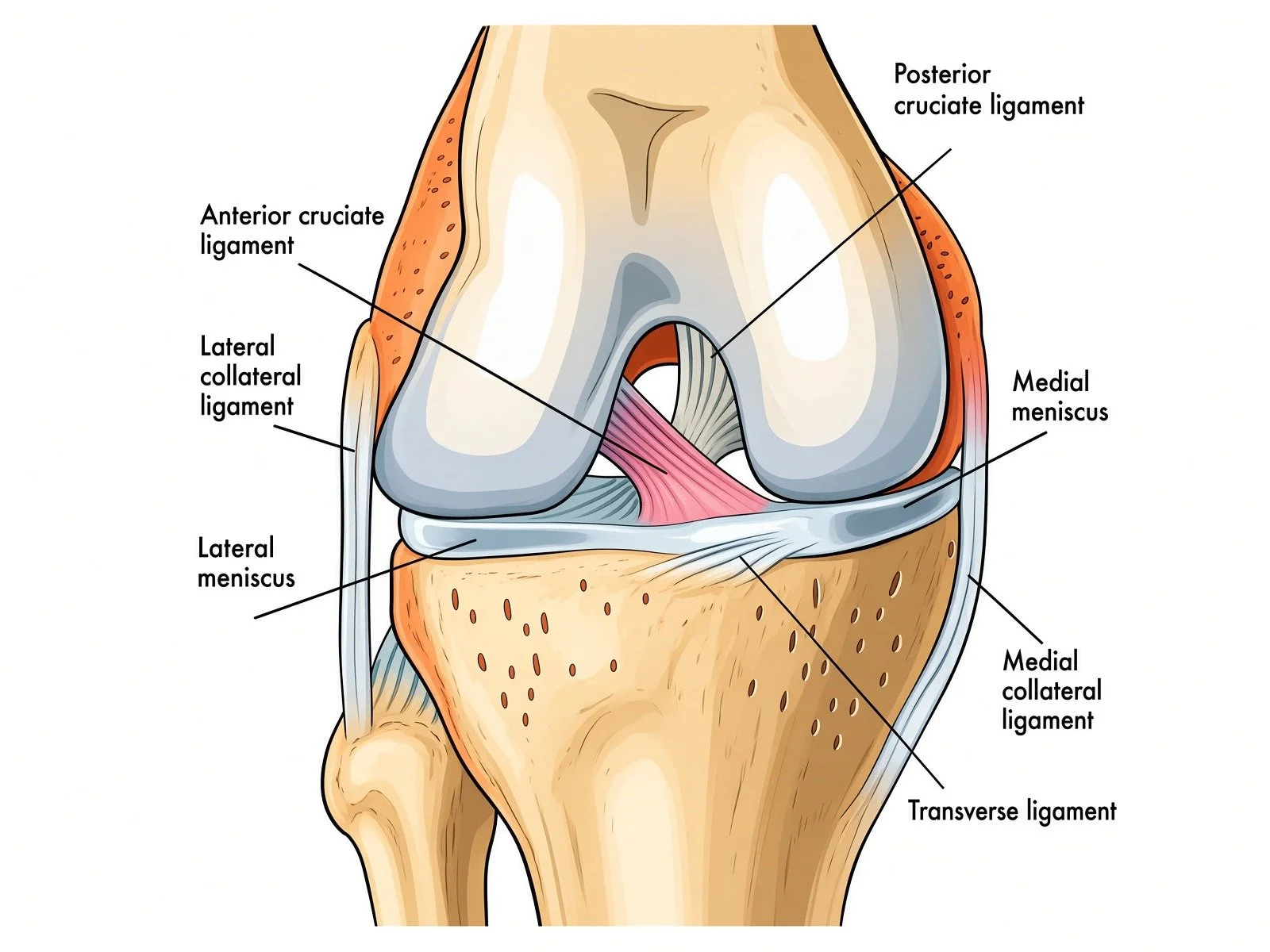
Where Is the ACL Located?
The ACL is located in the center of the knee joint, crossing in front of the posterior cruciate ligament (PCL)—which is how it gets its name, the anterior cruciate ligament.
It runs from the back portion of the femur to the front portion of the tibia, forming a strong connection between the two bones.
What Does the ACL Do?
Prevents the Tibia from Sliding Forward
Its primary function is to prevent the shinbone from moving excessively forward beneath the thighbone.
This is why physicians perform the Lachman test during an examination—the test directly evaluates this important function.
Controls Rotation of the Knee
Perhaps even more important for athletes, the ACL resists excessive rotation of the knee during activities such as:
Cutting
Pivoting
Changing direction
Landing from a jump
Sudden deceleration
These movements place tremendous forces across the ligament.
Provides Dynamic Stability
The ACL doesn’t work alone. It functions together with:
The meniscus
Other knee ligaments
The joint capsule
Surrounding muscles (especially the quadriceps and hamstrings)
When one structure is injured, the others often compensate—but only to a point.
Sends Important Signals to the Brain
One of the lesser-known functions of the ACL is proprioception—your body’s ability to sense where the knee is in space.
The ligament contains specialized nerve endings that constantly communicate with your brain, helping coordinate balance, agility, and reflexes. When the ACL tears, patients often describe their knee as feeling “untrustworthy” or “like it wants to give out.”
The Two Functional Bundles of the ACL
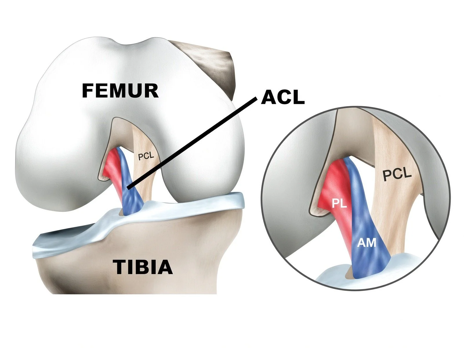
The ACL is made up of two distinct bundles of fibers that work together throughout the knee’s range of motion:
Anteromedial (AM) Bundle
Tighter when the knee is bent.
Plays a major role in controlling forward movement of the tibia.
Posterolateral (PL) Bundle
Tighter when the knee is straight.
Plays an important role in controlling rotational stability and preventing the knee from “giving way.”
Although these bundles function differently, they work together as a single ligament to provide stability throughout everyday activities and sports.
Signs of an ACL Tear
How does an ACL tear happen?
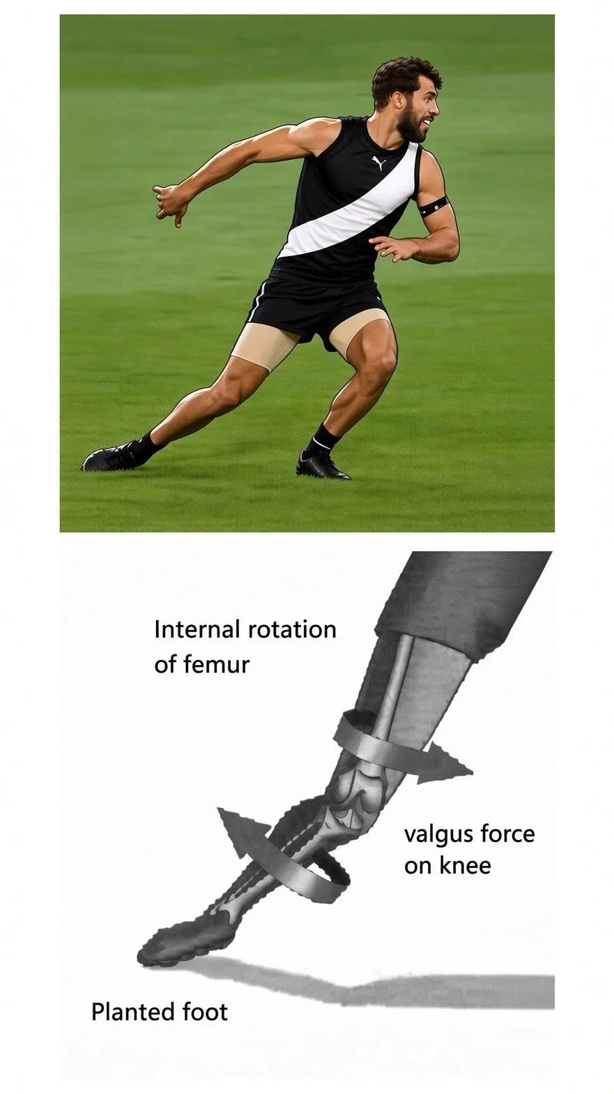
An ACL tear most commonly occurs during a sudden change in direction, pivot, or deceleration. Importantly, many ACL tears are noncontact injuries—there does not have to be another player or object striking the knee.
Common mechanisms include:
Pivoting or cutting: The foot is planted while the body suddenly changes direction.
Sudden deceleration: Slowing down rapidly from running or sprinting.
Awkward landing: Landing from a jump with the knee in a vulnerable position.
Hyperextension: The knee is forced beyond its normal range of motion.
Contact injury: A blow to the outside of the knee or another force can cause the knee to buckle or shift.
A classic ACL injury may feel like the knee “gave way” or shifted, sometimes accompanied by a pop. However, not everyone experiences or remembers a pop.
What are the symptoms of an ACL Tear?
Diagnosis of an ACL Tear starts with the history. With an ACL injury, patients may experience:
A pop or popping sensation at the time of injury
Sudden knee pain
Rapid swelling, often developing within the first several hours
Difficulty continuing to play or bear weight
A sensation that the knee gave way, buckled, or shifted
Loss of normal knee motion, particularly difficulty fully straightening the knee
Knee instability with subsequent cutting, pivoting, or athletic activity
Rapid swelling is particularly important. A knee that becomes significantly swollen shortly after an injury may have bleeding inside the joint (hemarthrosis). An ACL tear is one of the important causes of an acute traumatic hemarthrosis.
Physical Examination
Your orthopedic surgeon will first examine the knee for swelling, range of motion, tenderness, and stability.
Joint Effusion
An effusion means there is excess fluid inside the knee joint. After an acute injury, a significant effusion can be an important clue that something substantial has happened inside the knee.
Lachman Test
With the knee slightly bent, the examiner stabilizes the thigh while gently pulling the shin forward. Increased forward movement of the tibia compared with the opposite knee, particularly when there is a soft or absent endpoint, suggests ACL insufficiency.
Anterior Drawer Test
The anterior drawer test evaluates forward movement of the tibia relative to the femur with the knee bent approximately 90 degrees.
Increased anterior translation compared with the opposite knee can indicate an ACL tear. However, hamstring activation and other factors can make this test less reliable in the acutely injured knee.
Pivot Shift Test
The pivot shift test evaluates the functional instability caused by an ACL-deficient knee.
The examiner applies specific rotational and valgus forces while moving the knee through a range of motion. In an ACL-deficient knee, the tibia can shift relative to the femur, producing a characteristic “pivot shift.”
The pivot shift can be particularly useful for demonstrating rotational instability, although it can be uncomfortable in an acutely injured knee and is often easier to perform once swelling and pain have improved or when a patient is under anesthesia such as during surgery.
Imaging
1. X-Rays
Although X-rays do not directly show the ACL, they are an important part of the evaluation of a suspected ACL injury.
X-rays can:
Identify fractures or other bony injuries
Evaluate the overall alignment of the knee
Identify associated injuries that may occur with an ACL tear
Look for a Segond fracture
Evaluate the tibial slope, which can be relevant when assessing ACL injury risk and knee stability
What is a Segond Fracture?
A Segond fracture is a small bony avulsion involving the outer (lateral) portion of the proximal tibia.
It is thought to represent portions of the anterolateral ligament complex as well as posterior insertional fibers of the Iliotibial band (IT Band).
Although small, a Segond fracture is strongly associated with ACL injury and should raise suspicion for an ACL tear.
Tibial Slope
The posterior tibial slope can also be assessed on a lateral X-ray. A steeper posterior tibial slope has been associated with increased stress on the ACL and may be relevant when evaluating patients with ACL injuries, particularly those with recurrent instability or subsequent ACL tears
2. MRI
MRI is the most useful imaging study for confirming an ACL tear and evaluating the rest of the knee.
MRI can show:
Whether the ACL is intact, partially torn, or completely torn
The location and pattern of the ACL injury
Bone bruising associated with the injury
Meniscus tears
Cartilage injuries
Collateral ligament injuries
Other ligament injuries
Osteochondral injuries and fractures
MRI is particularly valuable because an ACL tear rarely occurs in isolation. Identifying associated injuries can influence treatment decisions and help your surgeon develop the appropriate treatment plan.
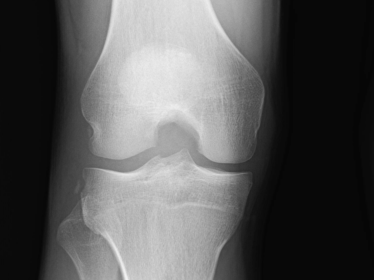
X-ray with a Segond Fracture
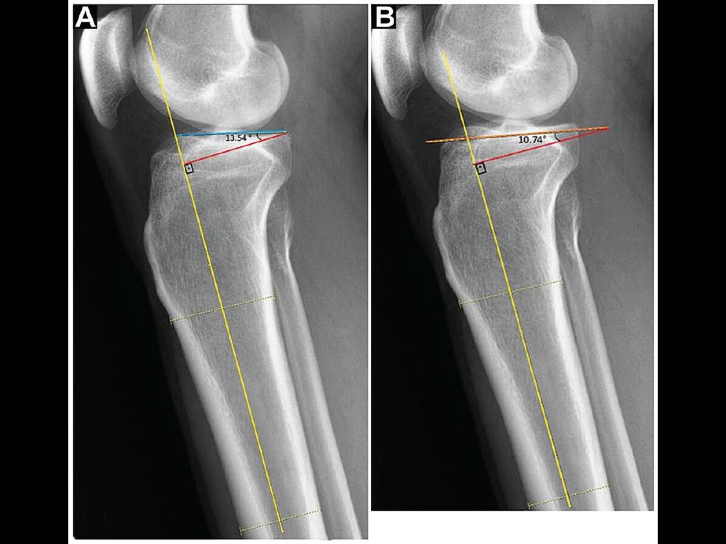
X-rays evaluating tibial slope
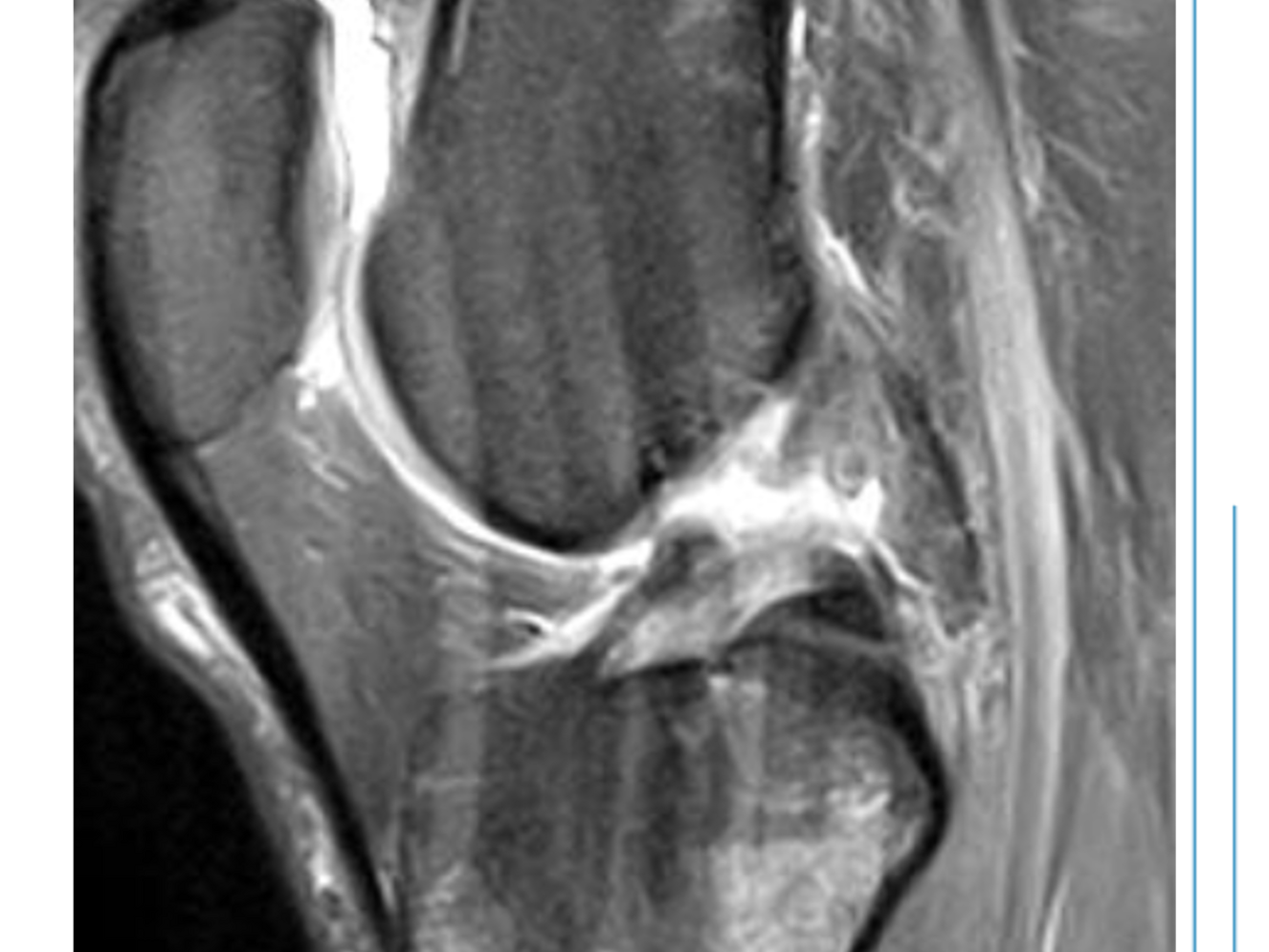
MRI of Midsubstance ACL Tear
Do I Need ACL Surgery?
Not everyone who tears their ACL needs surgery.
The decision to undergo ACL reconstruction depends on several factors, including your activity level, desired activities, age, symptoms, knee stability, associated injuries, and response to nonoperative treatment.
For some people, treatment without surgery can provide a stable, functional knee. For others—particularly athletes who want to return to cutting, pivoting, or high-demand sports—ACL reconstruction may provide the best opportunity to safely return to their desired activities and reduce the risk of ongoing instability.
Who Is More Likely to Benefit From ACL Surgery?
1. Your Activity Level and Desired Activities
One of the most important questions is not simply “How old are you?” but “What do you want your knee to be able to do?”
The ACL is particularly important for controlling the knee during activities involving rapid changes in direction and rotation.
If you want to return to activities such as: Soccer, Basketball, Football, Lacrosse, Tennis, Pickleball, Skiing or Snowboarding, Volleyball, Wrestling, Gymnastics Or other cutting, pivoting, or jumping sports, then ACL reconstruction may be recommended, particularly if your knee remains unstable.
In contrast, someone whose activities consist primarily of walking, cycling, swimming, strength training, or other activities that place less rotational demand on the knee may be able to function successfully without ACL reconstruction.
The goal is not simply to treat the MRI. The goal is to treat the patient and restore the level of function that is important to them.
2. Repeated Episodes of Instability or “Giving Way”
A knee that repeatedly gives way is an important reason to consider surgery.
Every episode of instability can place additional stress on other structures in the knee, including the meniscus and articular cartilage.
Some patients initially choose nonoperative treatment but later experience instability when they return to sports or other activities. If the knee continues to buckle despite appropriate rehabilitation and activity modification, ACL reconstruction may become the better option.
3. Failure of a Structured Nonoperative Treatment Program
Nonoperative treatment is a reasonable option for some patients.
A structured program may include:
Physical therapy
Regaining full knee motion
Reducing swelling
Restoring quadriceps and hamstring strength
Improving balance and neuromuscular control
Gradually returning to appropriate activities
Bracing
Some patients can achieve good function with this approach.
However, if you complete an appropriate rehabilitation program and continue to experience instability, giving-way episodes, or an inability to participate in the activities that are important to you, surgery may be recommended.
4. Your Age and Long-Term Goals
Age is an important part of the decision, but there is no single age at which ACL surgery is either required or inappropriate.
A younger athlete who wants to return to competitive pivoting sports may have a strong reason to consider reconstruction. At the same time, an active adult in their 40s, 50s, or beyond who wants to continue skiing, playing tennis, participating in recreational sports, or performing other high-demand activities may also benefit from reconstruction.
Age also matters because the risk of additional knee injury can change over time. Recurrent instability in an ACL-deficient knee can increase the likelihood of additional meniscus and cartilage damage and subsequent arthritis.
The decision should therefore consider both your current age and how you want to use your knee for the next several decades.
5. Associated Injuries That Require Surgery
Sometimes the ACL tear is only one part of the injury.
An associated injury may create a surgical indication even when the ACL itself might otherwise be treated without reconstruction.
Examples include:
Bucket-handle meniscus tear:
A displaced meniscus tear can cause the knee to lock or prevent the knee from fully straightening. Some of these tears are repairable and may require surgery.
Repairable meniscus injury:
Certain meniscus tears have a meaningful opportunity for healing or preservation through surgical repair. In an unstable ACL-deficient knee, treating the ACL and meniscus together may be recommended.
Cartilage or osteochondral injury:
Significant cartilage damage or an unstable cartilage/bone fragment may require surgical treatment.
Multiligament knee injury:
When an ACL tear occurs along with injuries to other major knee ligaments, such as the PCL or collateral ligaments, surgical treatment is more commonly necessary and requires individualized planning.
When Might Surgery Not Be Necessary?
Not every ACL tear requires reconstruction.
Nonoperative treatment may be reasonable when:
You do not participate in cutting or pivoting activities
You are comfortable modifying activities that make your knee unstable
Your knee is functionally stable during your desired activities
You have no recurrent episodes of giving way
You can regain good strength, motion, and function through rehabilitation
There are no associated injuries requiring surgery
Your goals can be achieved without reconstruction
Some patients with ACL tears become “copers”—people who are able to function well despite an ACL-deficient knee.
The Bottom Line
The decision to have ACL surgery should be based on your symptoms, your goals, your activity level, the stability of your knee, your age, and the condition of the rest of your knee—not simply the fact that your MRI shows an ACL tear.
If you want to return to cutting, pivoting, or high-demand sports and your knee remains unstable, ACL reconstruction is often recommended.
If your knee is stable and your desired activities do not require significant pivoting, a structured nonoperative program may be a reasonable alternative.
The right treatment is the treatment that gives you the best chance of maintaining a stable, functional knee for the activities you want to enjoy—both now and in the future.
ACL Surgery Basics
1. What Is the purpose of ACL Surgery?
The purpose is to restore functional stability to the knee, allowing the patient to return to desired activities and helping protect the meniscus and cartilage from recurrent instability.
Important distinction:
ACL surgery is not simply about “fixing the MRI.”
The goal is to restore functional stability.
There are different surgical approaches depending on the injury and patient: reconstruction, ACL repair, and BEAR.
Not every ACL tear requires surgery.
2. Arthroscopic Surgery
ACL surgery is typically performed using a minimally invasive arthroscopic technique. Small incisions allow a camera and specialized instruments to be placed into the knee, allowing the surgeon to directly visualize and treat the structures inside the joint. Larger incisions to take a graft or address extra-articular injuries may be required depending on the injury.
3. Reconstruction vs. Repair vs. BEAR
ACL Reconstruction - The torn ACL is replaced with a tendon graft
ACL Repair - The patient’s native ACL is repaired when the tear pattern and tissue quality are appropriate
BEAR (Bridge Enhanced ACL Restoration) - A bridge-enhanced approach designed to facilitate healing of the patients own ACL using a collagen scaffold and their blood.
4. What Happens to the Torn ACL?
For reconstruction, the torn ACL is removed and a graft (tissue from another location) is used to create a new ligament.
For repair/BEAR, the goal is different: preserving and facilitating healing of the patient’s native ACL when the injury is appropriate for those techniques.
5. What About Meniscus or Cartilage Damage?
ACL injuries frequently occur with other injuries, and the surgeon may treat these during the same operation.
Examples:
Meniscus repair (goal is to preserve meniscus tissue whenever possible)
Partial meniscectomy (removal of Meniscus tissue that is beyond repair)
Cartilage procedures
Additional ligament procedures in selected multiligament injuries
6. Anesthesia
General anesthesia is commonly used.
A regional nerve block may be recommended for postoperative pain control.
The anesthesia team evaluates each patient individually.
7. How Long Does ACL Surgery Take?
ACL surgery itself commonly takes approximately 1–2 hours, although the total operating time can vary depending on the surgical technique and whether additional procedures are required.
the patient will spend additional time in preoperative and recovery areas
8. Outpatient vs. Overnight
For uncomplicated ACL surgery, the vast majority of patients can expect outpatient surgery and to go home the same day. However, circumstances such as associated injuries, medical issues, pain control, or multiligament surgery can change this.
9. Braces, Crutches and Weight Bearing
postoperative restrictions vary depending on:
Surgical technique
Meniscus repair
Cartilage procedures
Associated ligament injuries
Surgeon-specific rehabilitation protocol
Some patients can bear weight relatively quickly, while others may require a period of restricted weight bearing. In general, patient’s with isolated ACL surgery and stable patterns of meniscus tears are able to weight bear immediately, but will typically still require crutches and braces for mobilizing.
10. When Does Rehabilitation Start?
Rehabilitation begins immediately after ACL surgery.
The early goals generally include:
Controlling swelling
Regaining full knee extension
Restoring knee flexion
Activating the quadriceps
Normalizing walking mechanics
Gradually restoring strength and neuromuscular control
11. How Long Until I Can Return to Sports?
Returning to sports after ACL surgery is based on biologic healing, strength, movement quality, psychological readiness, and functional testing—not simply the number of months since surgery..
return to cutting/pivoting sports generally takes many months (frequently 9 months or greater) and that returning too early can increase reinjury risk.
12. What Are the Risks?
Infection
Blood clots
Stiffness/loss of motion
Persistent pain
Weakness
Numbness around the incision and portions of the knee/leg
Meniscus/cartilage problems
Graft failure or recurrent ACL injury
Continued instability
Need for additional surgery
Complications related to anesthesia
Risks related to each individuals health or circumstances
13. Can an ACL Repair/Reconstruction Tear Again?
ACL reconstruction can provide excellent stability, but it does not make the knee immune to future injury. A repaired/reconstructed ACL can be reinjured, and some patients can also sustain an ACL tear in the opposite knee.
Age, activity level, return-to-sport timing, neuromuscular control, and participation in high-risk sports all influence reinjury risk.
ACL Reconstruction & Graft Selection
ACL Reconstruction: What Happens During Surgery?
ACL reconstruction is typically performed arthroscopically through several small incisions. The exact procedure varies depending on the graft being used and whether there are associated meniscus, cartilage, or ligament injuries.
1. Examination of the Knee
After anesthesia is administered, your surgeon examines the knee and compares its stability with the opposite knee.
This allows the surgeon to confirm the degree of ACL instability and evaluate the knee before beginning the reconstruction.
2. Arthroscopic Evaluation
A small camera called an arthroscope is placed into the knee through a small incision.
This is important because ACL injuries can occur along with meniscus tears, cartilage injuries, or other ligament injuries.
3. Treating Associated Injuries
If another injury is identified, it may be treated during the same operation.
Whenever possible, meniscus tissue is preserved and repaired rather than removed, particularly when the tear has a reasonable chance of healing.
Other associated injuries may also require treatment depending on their location, severity, and stability.
4. Preparing the ACL Graft
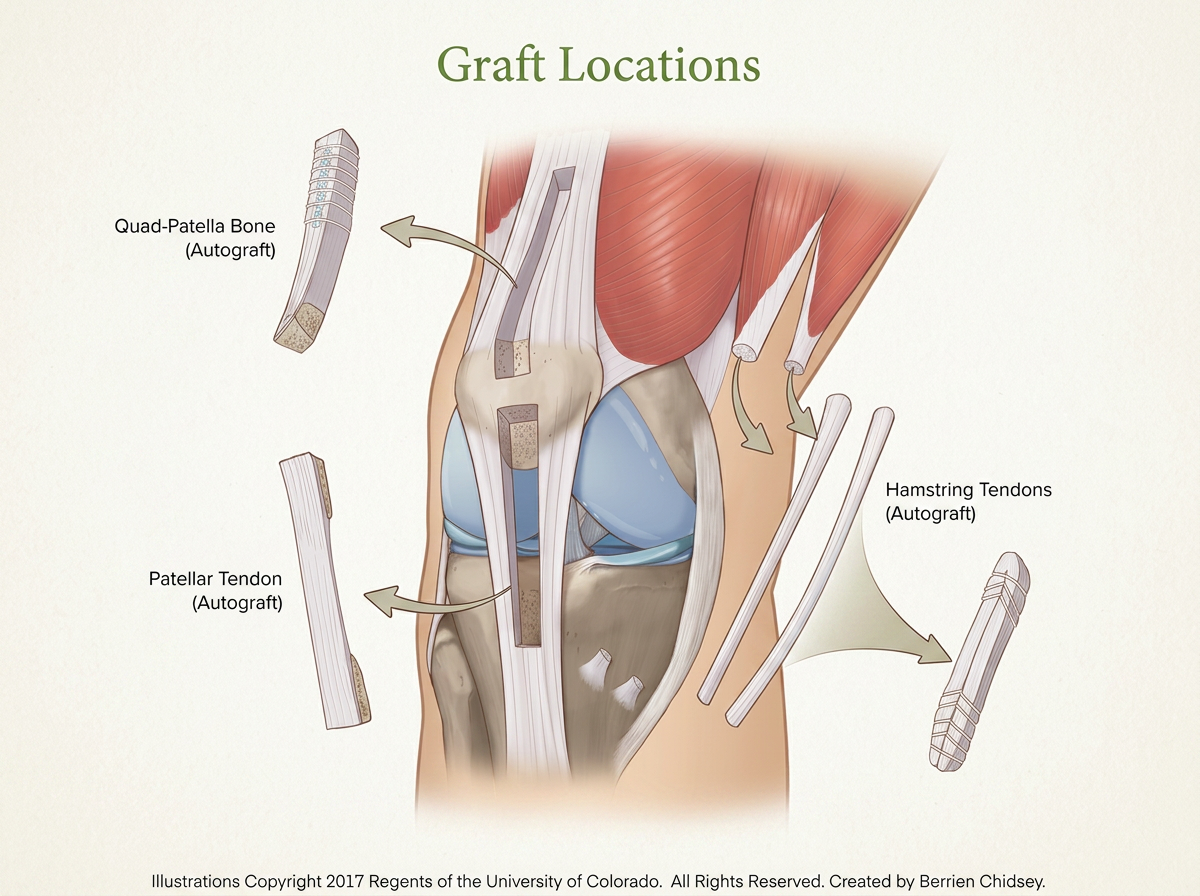
For an ACL reconstruction, a tendon graft is used to create a new ACL.
Depending on the graft selected, the tissue may come from:
The patient’s quadriceps tendon
The patient’s patellar tendon
The patient’s hamstring tendons
Donor tissue (allograft)
The graft is carefully prepared to the appropriate size and shape for the patient’s knee.
5. Preparing the ACL Attachment Sites
The torn ACL is addressed arthroscopically, and the surgeon prepares the areas on the femur and tibia where the ACL normally attaches.
6. Creating the Bone Tunnels
Small tunnels are created in the femur and tibia at the appropriate ACL attachment locations.
These tunnels allow the new graft to be positioned where the native ACL was located.
The size and position of the tunnels are carefully determined based on the patient’s anatomy and the graft being used.
7. Positioning the Graft
The prepared graft is passed through the tunnels and positioned across the knee.
8. Securing the Graft
The graft is secured within the femur and tibia using specialized fixation devices.
The fixation method depends on the type of graft and the surgeon’s technique.
The surgeon then checks the graft’s position, tension, and stability through a range of motion.
9. Final Examination
Before completing the operation, the surgeon confirms that:
The graft is appropriately positioned
The knee has regained stability
The graft moves appropriately through the range of motion
There is no abnormal impingement
Any associated injuries have been appropriately addressed
10. Closing the Incisions
The instruments are removed, the small incisions are closed, and a dressing is placed over the knee.
The patient then goes to the recovery area where the anesthesia team monitors their recovery.
Autograft
Patient’s own tissue
Generally preferred for young/high-demand athletes because of lower failure/retear rates compared with allograft
Requires harvesting tissue from the patient
Donor-site morbidity: term used to describe possible pain/problems that can occur from the graft harvest site.
Biological incorporation is generally favorable
Most common options: quadriceps tendon, patellar tendon, hamstring
Allograft
Donor tissue from a tissue bank
No harvest-site morbidity
Avoids creating another surgical site in the patient’s leg
However, particularly in young/high-demand patients, has higher reported failure rates than autograft
Incorporation/remodeling is generally slower
Very low but nonzero risk of disease transmission despite modern tissue-bank screening/processing
Particularly useful in selected older/less active patients, revision situations, or when autograft tissue is inadequate
Bone Patellar Tendon Bone
BTB has excellent stability but comes with a more recognizable anterior-knee donor-site morbidity profile.
Advantages:
Excellent fixation with bone-to-bone healing
Long track record
Particularly well established in high-level athletes
Low failure rates
Potential Disadvantages:
Anterior knee pain
Pain with kneeling
Patellar tendon symptoms
Risk of patellar fracture
Risk of tibial tubercle fracture
Potential extensor mechanism complications
Numbness around the incision
Hamstring
Advantages:
Less anterior knee morbidity than BTB
Relatively small harvest incision
Familiar, well-established technique
Potential Disadvantages:
Hamstring weakness
Potential deficits in deep knee flexion and dynamic stabilization
Tendon regeneration is variable
Graft size can vary
Smaller grafts may be associated with increased failure risk
Some comparison studies demonstrating higher retear risk compared to other autograft options.
Key Concept with Autografts:
Every autograft has a trade-off: you are using your own tissue to reconstruct the ACL, but that tissue has to come from somewhere.
Quadriceps Tendon
A strong, versatile graft that has become increasingly popular for ACL reconstruction.
Advantages:
Large cross-sectional area
High graft strength
Can provide substantial graft volume - predictable graft size
Can be harvested with or without a bone block depending on technique
Generally less anterior knee morbidity/pain than BTB
Particularly attractive for athletes and active patients
Useful in primary and some revision reconstructions
Potential Disadvantages:
Anterior thigh/knee pain can occur
Temporary quadriceps weakness is expected, rarely permanent
Quadriceps strength recovery can be an important component of rehabilitation
Numbness or sensitivity around the harvest site
Risk of tendon-related complications, although uncommon
Technique and graft sizing matter
*For many of my patients, I favor quadriceps tendon autograft because it provides an excellent combination of graft size and strength while avoiding some of the donor-site concerns associated with other commonly used grafts. However, I do not believe one graft is right for every patient.
The BEAR® Implant:
Helping Your ACL Heal Instead of Replacing It
For many years, an ACL tear meant one thing: reconstruction. The torn ACL was removed or trimmed, and a tendon from another part of the body was used to create a new ligament. But there is another possibility for appropriately selected patients:
What if we could help your own ACL heal?
That is the idea behind the BEAR® (Bridge-Enhanced ACL Restoration) Implant.
Instead of removing the torn ACL and replacing it with a tendon graft, the BEAR procedure uses a collagen-based implant and your own blood to create an environment that allows the torn ends of your native ACL to heal together.
Why Doesn’t the ACL Normally Heal on Its Own?
The ACL is different from many other ligaments in the body.
When you cut your skin or tear a ligament outside of a joint, bleeding occurs and a blood clot forms between the injured ends. That clot acts as a temporary scaffold. It contains cells and growth factors that help organize the healing process.
The ACL, however, sits inside the knee joint and is surrounded by synovial fluid.
When the ACL tears, the initial blood clot and the biological signals needed for healing can be disrupted by the constant flow of joint fluid. The injured ligament is also exposed to a biologically active environment containing enzymes that can break down the proteins and extracellular matrix that are necessary for ligament healing.
In other words: The problem isn’t necessarily that the ACL doesn’t have the ability to heal. The problem is that the environment inside the knee makes it very difficult for the normal healing process to occur.
How Does the BEAR Implant Change This?
The BEAR Implant is designed to change the biological environment around the torn ACL.
During the procedure, the torn ACL is repaired and the BEAR Implant is placed between and around the torn portions of the ligament. A small amount of the patient’s own blood is added to the implant.
The collagen implant acts as a temporary biological bridge and scaffold. Think of it as a cocoon to allow healing to take place.
It helps:
Hold the patient’s blood and developing clot at the site of the ACL tear
Provide a framework for new tissue formation
Bring the torn ACL ends together
Support the body’s natural healing response
Preserve the patient’s remaining native ACL tissu
As healing progresses, the implant is gradually resorbed and replaced by newly formed ligament tissue.
So rather than:
Torn ACL → remove it → replace it with a tendon graft
the goal is:
Torn ACL → support the healing environment → allow the patient’s own ACL to restore itself
The BEAR procedure combines repair of the ACL with a collagen-based scaffold and the patient’s own blood to facilitate restoration of the native ligament. Clinical studies have demonstrated outcomes that are non-inferior to hamstring autograft reconstruction at two years, with better preservation of hamstring strength. (PubMed)
Where Does BEAR Have an Advantage?
1. It Preserves Your Native ACL
One of the most important differences between BEAR and ACL reconstruction is that we are trying to preserve your ACL rather than replace it.
Your native ACL contains its original attachments, blood supply, and sensory nerve fibers involved in proprioception—the ability of your brain to sense the position and movement of your knee.
This can be particularly important in a partial ACL tear, where a significant amount of healthy ACL tissue may still be present.
With conventional reconstruction, some of this remaining tissue generally has to be removed to make room for the graft. Research has demonstrated proprioceptive nerve fibers within ACL remnants even years after injury.
With BEAR, the goal is to preserve and restore as much of that native tissue as possible.
3. Potentially Lower Risk of Post-Traumatic Arthritis
ACL surgery isn’t just about getting an athlete back on the field. The knee has to last for decades.
In the BEAR I and BEAR II studies, patients treated with BEAR had a substantially lower rate of radiographically confirmed post-traumatic osteoarthritis at six years compared with patients who underwent ACL reconstruction using hamstring autograft.
Six times Lower rate of radiographically confirmed post-traumatic osteoarthritis
The difference was statistically significant, and in January 2026 the FDA updated the BEAR Implant’s labeling to include this reduced risk of post-traumatic osteoarthritis in appropriately indicated patients. (Business Wire)
Our goal should be to give you a stable knee today—and preserve that knee for the rest of your life.
2. BEAR for Partial ACL Tears
Partial ACL tears represent a particularly challenging dilemma, as a reconstruction would involve removal of substantial ACL tissue.
14-56% of partial tears will progress to full tears (2, 3)
As high as 86% Progression if bone marrow edema on MRI
5.2x higher likelihood of progression in patients less than 20 years of age
50% of those that progress will have a meniscus tear
Addition of meniscus tear increases likelihood of development of posttraumatic arthritis.
Only 44% of those with a partial ACL tear are able to return to preinjury level of sports. (2)
A subset of BEAR III Trial clinical data at 2 years demonstrated a 0% retreat rate amongst 27 patients.
4. No Tendon Graft Harvest
Traditional ACL reconstruction requires obtaining a tendon graft.
This creates an additional surgical site and can produce what is known as donor-site morbidity.
Potential problems vary depending on the graft but can include:
Hamstring weakness
Anterior knee pain
Kneeling discomfort
Quadriceps weakness
Altered sensation around the harvest site
Additional postoperative pain
BEAR does not require harvesting a tendon from another part of your body.
Your ACL is the graft.
Physical Therapy & Rehabilitation
Surgery Repairs the ACL. Rehabilitation Rebuilds the Knee.
ACL surgery is only the beginning of your recovery.
The surgery reconstructs the torn ligament, but rehabilitation is what helps you regain:
Full knee motion
Quadriceps strength
Normal walking
Balance and coordination
Confidence in your knee
The ability to run, jump, cut, and change direction safely
A technically excellent ACL reconstruction can still result in a stiff, weak, or poorly functioning knee if rehabilitation is not done consistently and appropriately.
Your rehabilitation is an active part of your ACL treatment—not something you do after treatment.
Most patients should expect a gradual recovery over many months. Returning to unrestricted pivoting sports commonly takes 9–12 months or longer, and the decision to return should be based on how the knee is functioning rather than simply how much time has passed.
First Priority: Maintain/Get the Knee Straight
Full Extension Matters:
One of the most important goals immediately after ACL surgery is to regain full knee extension—being able to completely straighten your knee.
This deserves special emphasis because loss of extension is one of the problems we see most often after ACL reconstruction. It can mean returning to the OR!
A knee that remains slightly bent may not seem like a major problem at first. However, persistent loss of extension can contribute to:
A limp
Difficulty walking normally
Quadriceps weakness
Increased anterior knee pain
Difficulty progressing through rehabilitation
Ongoing stiffness
Reduced satisfaction with the outcome
It is much easier to prevent loss of extension early than to try to regain it months later.
Your goal is generally to regain the same amount of extension that you had before surgery. This is important because some people naturally have a small amount of hyperextension. Your “normal” may not be the same as someone else’s.
Do not routinely place a pillow directly underneath your knee while resting.
Although this position may feel comfortable, keeping the knee bent for prolonged periods can make it harder to regain full extension.
Instead, when your goal is to work on extension, support the heel or ankle and allow the knee to straighten.
Tell us if your knee is not getting straight
Do not assume that loss of extension is something you simply need to “work through.”
If your knee is not progressively straightening during the first several weeks—or if you feel that your progress has stopped—tell your surgeon and physical therapist.
Early recognition of an extension problem is important
Quad Sets
Tighten the thigh muscle and press the back of your knee toward the surface underneath you.
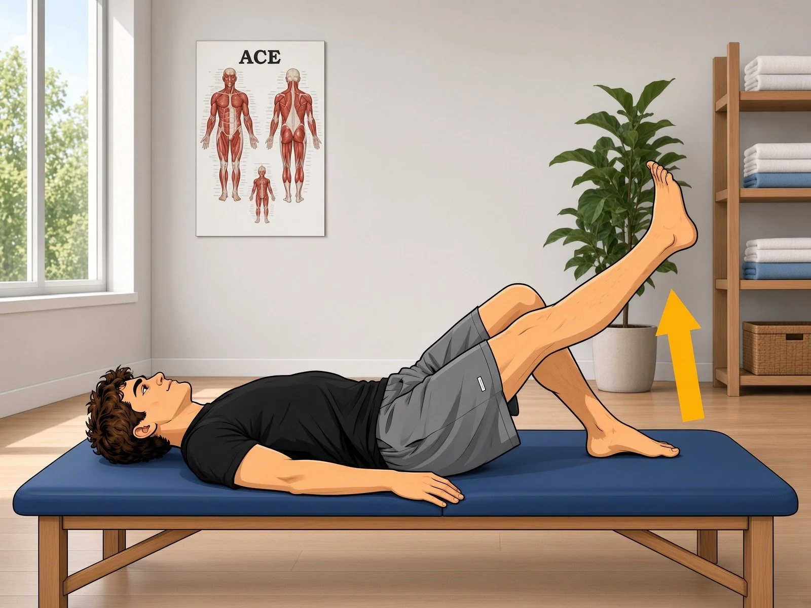
Straight Leg Raises
Once you can adequately activate the quadriceps and perform the movement safely, straight-leg raises help reinforce quadriceps control.
Phase 3: Build Strength
Approximately 3–6 months
This is where rehabilitation starts to look much more like athletic training.
The focus shifts toward developing meaningful strength in the surgical leg and restoring control during more demanding movements.
Your therapist may progress:
Squats
Split squats
Step-downs
Single-leg strengthening
Leg press
Hamstring strengthening
Calf strengthening
Hip strengthening
Cardiovascular conditioning
Running is not automatically started because you have reached a particular month after surgery.
Before running, you should demonstrate adequate strength, motion, control, and minimal swelling.
Your therapist may use strength testing and movement assessment to determine whether your knee is ready.
Phase 5: Return to Sport
Approximately 9–12+ months
Returning to sports that involve cutting, pivoting, jumping, or rapid changes in direction is the final stage of rehabilitation.
But time alone should not determine when you return to sport.
You may feel good at six months and still not be ready.
Conversely, another patient may progress faster or slower depending on their individual circumstances.
Your Home Program Matters
Physical therapy does not only happen in the therapy clinic. Your home program is an important part of your recovery.
Patients who tend to progress best aren’t necessarily those who perform the hardest exercises. Often they are the most consistent
Strategies for Maintaining Extension
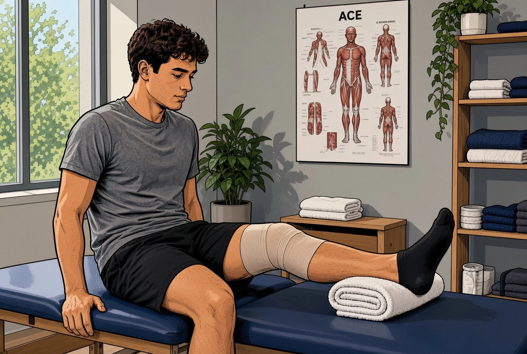
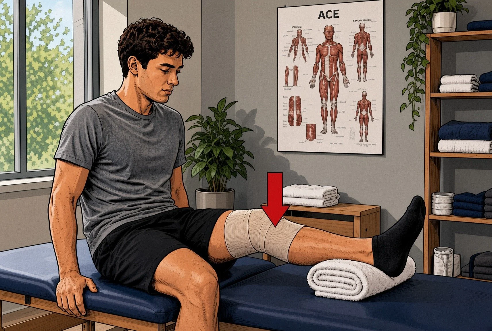
Heel-prop exercises
Place your heel on a pillow, rolled towel, or other support so that your calf and thigh are unsupported. Allow gravity to gently straighten the knee.
Quadriceps activation
A functioning quadriceps helps keep the knee straight. Contracting the quadriceps and attempting to push the knee into the table aids with return of extension.
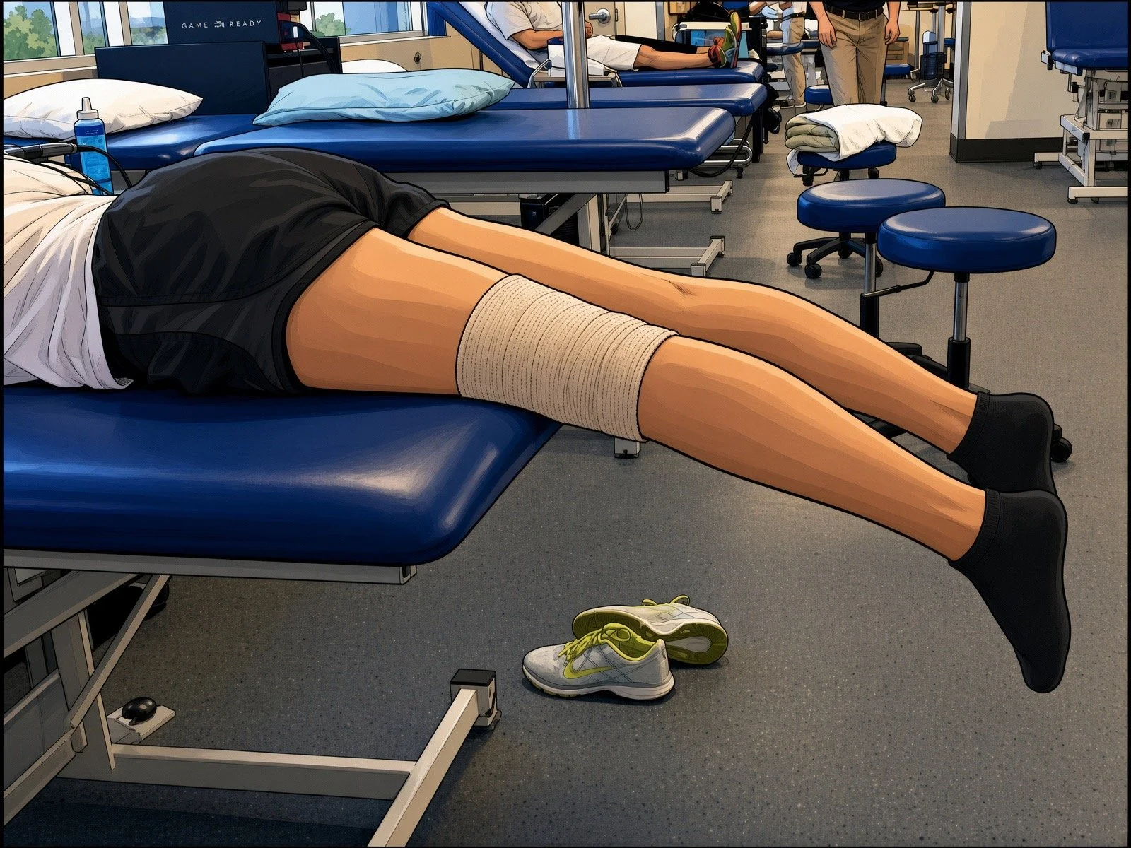
Prone Knee Hangs
Laying flat on your stomach (prone) with knees hanging over the edge of a table or bed and allowing gravity to pull the knee into a straight position.
Controlling Swelling
Swelling is normal after ACL surgery, but excessive swelling can interfere with almost every aspect of rehabilitation.
A swollen knee is harder to move, can limit return of motion, and swelling can inhibit the quadriceps muscle. This creates a cycle:
Swelling → poor quadriceps activation → weakness → difficulty walking → slower recovery
For this reason, swelling control is not simply about making your knee feel better. It is part of restoring normal knee function.
Common strategies include:
Ice or cold therapy
Compression
Elevation
Gentle motion
Appropriate activity levels
Following your postoperative instructions
Your knee does not need to be completely pain-free or swelling-free before you begin rehabilitation. However, increasing swelling after exercise is often a sign that you are doing more than your knee is currently ready to handle.
Wake Up Your Quadriceps
Your Thigh Muscle May “Shut Down”
The quadriceps muscle is one of the most important muscles for ACL recovery.
After surgery, it is common for the quadriceps to become surprisingly difficult to activate. This is partly due to pain and swelling and partly due to a phenomenon called arthrogenic muscle inhibition, in which the nervous system essentially decreases the muscle’s ability to fully contract.
You may feel like you are trying to tighten your thigh but simply cannot make it work normally.
This is common—and it is something we actively work to overcome.
Early quadriceps exercises may include:
Quad Sets
Straight-Leg Raises
Electrical Muscle Stimulation (NMES)
Your therapist may use electrical stimulation to help recruit the quadriceps when voluntary activation is difficult.
Biofeedback
Some therapists use visual or auditory feedback to help you understand when and how strongly your quadriceps is contracting.
The goal is not simply to perform these exercises.
The goal is to teach your brain and quadriceps to work together normally again.
As your knee improves, rehabilitation progresses from simple activation exercises to progressively heavier strengthening.
Bracing and Crutches
Protecting the Knee While You Learn to Walk Again
After surgery, we typically have patients use a T-scope-style hinged knee brace during the early recovery period.
The brace provides protection while the reconstructed ACL and any additional repairs are healing. It can also provide additional support while your quadriceps is still weak.
Your exact brace settings and how long you need to use it will depend on your surgery and your progress.
Do not change the brace settings or discontinue the brace simply because the knee feels better. Follow the instructions provided by your surgical and rehabilitation team.
Crutches
Crutches are used to protect the knee and help you develop a safe walking pattern while your strength and control return.
The goal is not simply to “get off the crutches.” The goal is to walk normally without them.
If you stop using crutches while you are still limping, you may simply teach yourself to walk with an abnormal pattern.
We want to see:
The knee able to fully straighten during walking
Appropriate heel-to-toe progression
Good quadriceps control
Minimal or controlled swelling
No significant limp
Appropriate weight acceptance through the surgical leg
Once those criteria are met, you can progressively transition away from the crutches according to your therapist’s and surgeon’s instructions.
The Rehabilitation Journey
Your rehabilitation should progress according to what your knee can do, not simply according to the calendar.
The exact timeline will vary depending on:
The type of ACL reconstruction
Your graft choice
Meniscus repair or other procedures performed
Cartilage treatment
Your age and activity level
Your strength before surgery
Your individual healing response
The following is a general framework:
Phase 1: Protect the Knee and Restore Motion
Surgery through approximately 6 weeks
The early goals are:
1. Control pain and swelling
Use ice, compression, elevation, and appropriate activity.
2. Regain full extension
This is one of the highest priorities of the entire early phase.
3. Restore bending
Gradually work toward your normal range of motion as permitted by your surgeon and therapist.
4. Wake up the quadriceps
Begin with activation exercises and progress as your muscle control improves.
5. Learn to walk normally
Use the brace and crutches as instructed, with the goal of restoring a normal walking pattern before abandoning them.
6. Protect any additional surgical repairs
If you have a meniscus repair, cartilage procedure, or other procedure performed at the time of ACL reconstruction, your early rehabilitation may be more restricted.
Phase 2: Restore Normal Motion/Build a Foundation
Approximately 6–12 weeks
Goals include:
Full extension equal to the opposite knee
Progress toward full flexion
Minimal swelling
Normal walking
Improving quadriceps strength
Improving hamstring and hip strength
Balance and proprioception
Progressively increasing cardiovascular fitness
Exercises become progressively more challenging, including movements such as:
Squats
Step-ups
Leg press
Bridges
Lunges when appropriate
Single-leg balance
Hip and core strengthening
The exact exercises and timing depend on your individual surgery and rehabilitation plan.
Phase 4: Running, Jumping, and Agility
Approximately 4–9 months
As strength and movement quality improve, rehabilitation progresses toward higher-level activities.
This may include:
Running
Acceleration and deceleration
Jumping and Landing mechanics
Hopping
Cutting
Agility
Change-of-direction drills
Sport-specific movements
The emphasis is not simply on whether you can perform a movement. We also want to know how you perform it. For example, we want to identify and correct:
Excessive knee collapse
Poor landing mechanics
Asymmetry
Poor trunk control
Inadequate hip control
Inability to absorb force appropriately
These movement patterns matter because returning to sport with poor mechanics may increase the stress placed on the reconstructed knee.
Don’t Chase the Calendar
It is tempting to ask:
“When can I run?”
“When can I play?”
“When can I stop going to therapy?”
Those are reasonable questions, but the better question is:
“What does my knee need to be able to do before I safely progress?”
ACL rehabilitation is increasingly based on criteria rather than time alone.
Two patients who are both four months from surgery may be at very different stages of recovery.
Your rehabilitation should be individualized to your knee, your surgery, your goals, and your progress.
The Bottom Line
ACL rehabilitation is a long-term process.
The early goals are simple but extremely important. From there, rehabilitation progresses toward running, jumping, cutting, sport-specific training, and ultimately a safe return to your desired activities.
There is no shortcut.
The surgery reconstructs the ligament. Your rehabilitation helps rebuild the knee.
And the goal is not simply to get back to doing what you were doing before your ACL injury.
The goal is to return with a knee that is strong, stable, functional, and prepared for the demands you are going to place on it.
Meniscus, Cartilage, and Other Injuries Associated With an ACL Tear
An ACL tear is often not the only injury inside the knee.
When the ACL tears, the same force that damages the ligament can also injure the meniscus, cartilage, or other ligaments. In addition, once the ACL is no longer functioning normally, the knee can continue to shift and rotate abnormally, potentially causing additional damage over time.
This is one reason that ACL treatment is not exactly the same for every patient.
Meniscus Tears
The meniscus is a C-shaped piece of cartilage that sits between the thighbone and shinbone. Each knee has a medial meniscus on the inside and a lateral meniscus on the outside.
The menisci are important because they:
Help distribute forces across the knee
Provide shock absorption
Improve knee stability
Help protect the articular cartilage
Contribute to normal knee motion
Meniscus tears are very common with ACL injuries. Depending on the patient population and the timing of surgery, studies have found meniscal injuries in roughly 20–65% of patients undergoing ACL reconstruction, with some studies reporting even higher rates in particular groups.
Why does the type of meniscus tear matter?
Not all meniscus tears need to be repaired.
Some tears are stable and can safely be left alone while the ACL is reconstructed. Others require repair because they can interfere with knee function or because preserving the meniscus is important for the long-term health of the knee.
Some meniscus injuries can also be difficult to identify on an MRI. During ACL surgery, we carefully inspect the menisci to look for injuries that may not have been obvious on the original imaging.
How Does Meniscus Surgery Affect ACL Recovery?
After a meniscus repair, your surgeon and physical therapist may temporarily restrict certain activities—such as deep bending, weight bearing, or loaded squatting—depending on the type and location of the repair.
This does not necessarily mean that your ACL is healing more slowly. Rather, the rehabilitation program must protect the repaired meniscus while the ACL graft is also healing.
As a result, two patients who undergo ACL reconstruction on the same day may have very different early rehabilitation programs depending on whether a meniscus was repaired.
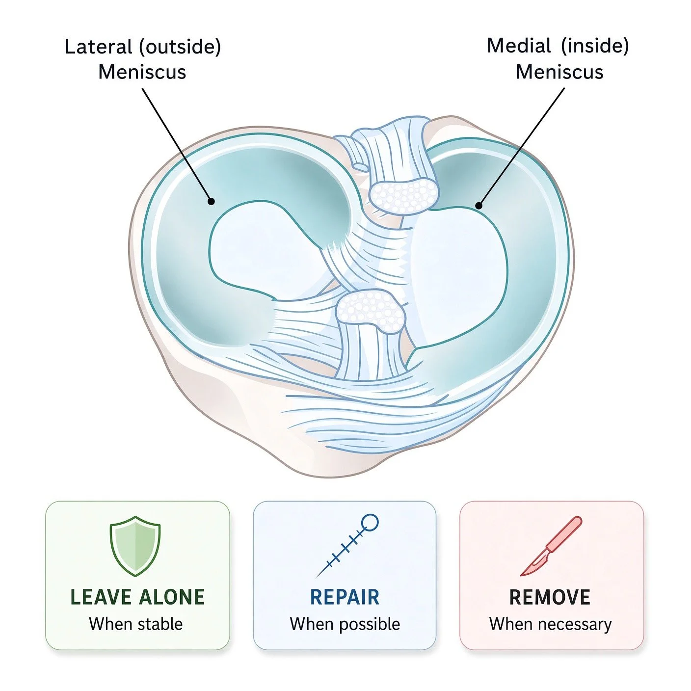
Options for Meniscus Tears:
1. Meniscus repair
The torn meniscus is sewn back together in an attempt to preserve as much normal tissue as possible.
Whenever it is reasonable, we favor meniscus preservation.
The meniscus acts as an important secondary stabilizer and helps protect the articular cartilage. Preserving a functional meniscus may therefore be important not only for your recovery from ACL surgery but also for the long-term health of your knee.
2. Partial meniscectomy
If a portion of the meniscus is badly damaged or cannot be repaired, the unstable portion may be removed.
Recovery from a partial meniscectomy is generally faster than recovery from a meniscus repair. However, removing meniscal tissue reduces the knee’s ability to distribute forces, which is one reason surgeons try to preserve the meniscus whenever possible.
3. Leaving a stable tear alone
In selected cases, a stable tear can be left in place while the ACL is reconstructed. This avoids unnecessary surgery and preserves the maximum amount of native meniscus.
Cartilage Injuries
The smooth surface covering the ends of the bones inside your knee is called articular cartilage.
Healthy cartilage allows the bones to glide smoothly and helps distribute forces across the knee.
Cartilage damage is also common with ACL injuries. More than one in four patients undergoing ACL reconstruction may have some degree of cartilage injury identified at surgery.
The treatment depends on:
The size of the defect
The depth of the injury
Its location
Whether the underlying bone is involved
Your age and activity level
The condition of the surrounding cartilage
Whether there is associated meniscus damage or knee instability
Chondroplasty
For smaller or superficial areas of damaged cartilage, the surgeon may smooth unstable cartilage and remove loose or damaged tissue.
The goal is to create a stable cartilage surface rather than attempting to replace the entire defect.
Cartilage Restoration Procedures:
Microfracture - small, full thickness defects treated by creating holes in the underlying bone to allow marrow elements to enter the defect and stimulate the formation of new cartilage.
OATS (Osteochondral Autograft Transfer - uses a small plug of healthy cartilage and underlying bone taken from another area of the knee and transfers it into the damaged area.
Osteochondral Allofraft - for larger defects, uses donor cartilage and bone to restore larger damaged areas.
MACI (Matrix-associated Autologous Chondrocyte Implantation - Donor cells harvested from patients knee and used to regrow cartilage in a lab, that must then later be implanted at a second surgery.
Additional Emerging Options
Other Ligament Injuries
The ACL is only one of four major ligaments of the knee.
An ACL tear can occur together with injuries to other ligaments, including:
MCL — the ligament on the inside of the knee
PCL — the ligament that helps prevent the tibia from moving too far backward
LCL and posterolateral corner — structures on the outside/back of the knee that help control stability and rotation
Treatment depends on the specific combination of injuries.
Some MCL injuries, for example, can heal without surgery while the ACL is reconstructed later. Other ligament injuries may require repair or reconstruction, and occasionally treatment is staged rather than performed all at once.
These injuries are particularly important because untreated instability in another part of the knee can place additional stress on an ACL reconstruction.
Lateral Extra-Articular Tenodesis
Some patients have more rotational instability than can be adequately controlled with an ACL reconstruction alone.
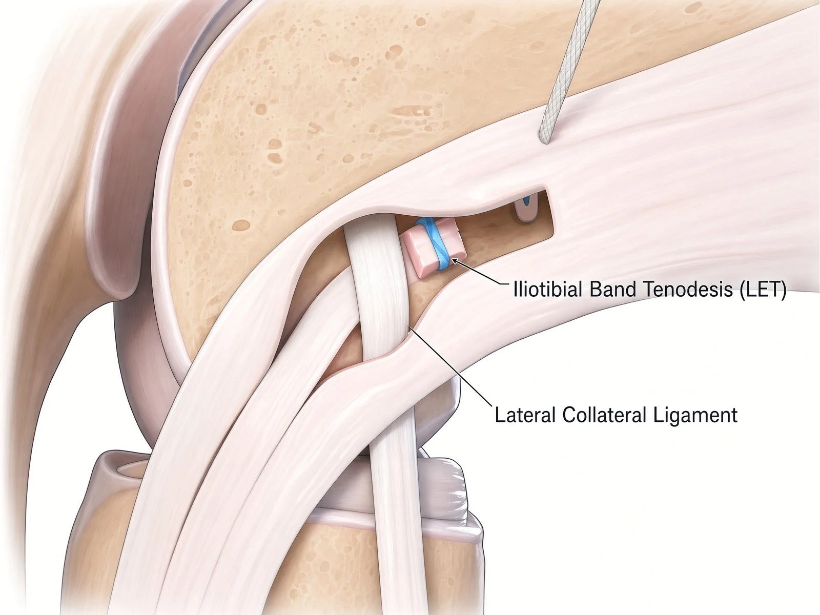
This is where a procedure called a lateral extra-articular tenodesis, or LET, may be helpful. Also referred to as Iliotibial Band Tenodesis
LET is an additional procedure performed on the outside of the knee. It works together with the ACL graft to help control excessive rotation of the tibia.
Think of the ACL reconstruction as restoring the knee’s primary internal restraint, while the LET provides an additional check against excessive rotational movement.
Who Might Benefit From LET?
LET is not necessary for every ACL reconstruction.
It is generally considered when the risk of graft failure or persistent rotational instability is higher than average.
Examples may include:
A high-grade pivot shift on examination (commonly this is determined during examination under anesthesia)
Young athletes returning to high-risk pivoting or cutting sports (less than 25 is a relative consideration)
Revision ACL reconstruction
Generalized ligamentous laxity
Certain patients with significant knee hyperextension
Selected patients with other factors that increase the risk of ACL graft failure
The decision is individualized based on your examination, activity goals, graft choice, and other characteristics of your knee.
A major randomized clinical trial involving young, high-risk patients found that adding LET to hamstring ACL reconstruction reduced graft rupture from approximately 11% to 4% over two years. (PubMed)
Other systematic reviews have similarly found lower graft-failure rates and less rotational instability when LET is added in appropriately selected high-risk patients. (PubMed)
LET can therefore be an important tool for reducing the risk of ACL reconstruction failure in the right patient—but it is not a routine addition to every ACL surgery.
Return to Sports
When can I return to sports?
One of the most common questions after ACL surgery is: “When can I play again?”
The better question is: “When is my knee, my strength, and my body ready for the demands of my sport?”
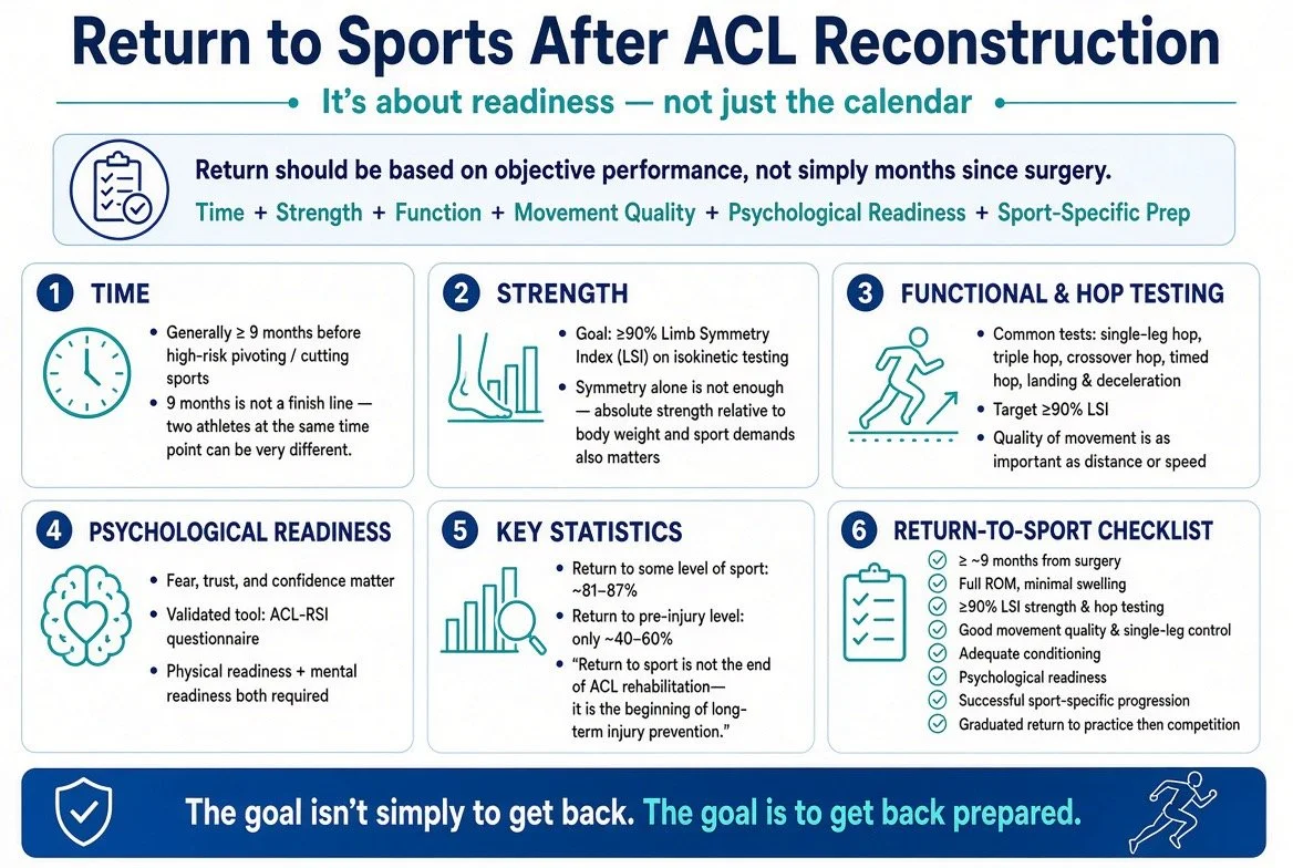
Returning to sports after ACL reconstruction should be based on objective performance and readiness—not simply the number of months since surgery.
Time is important because the reconstructed ACL needs time to biologically incorporate and mature. For athletes returning to sports that involve sprinting, jumping, cutting, pivoting, or contact, returning before approximately 9 months is generally discouraged.
But 9 months is not a finish line.
Two athletes can be 9 months from surgery and have very different levels of strength, movement quality, conditioning, confidence, and sport-specific readiness.
Think criteria—not calendar
A successful return to sport combines several pieces:
Time + Strength + Functional Performance + Movement Quality + Psychological Readiness + Sport-Specific Preparation
No single test can determine whether you are ready.
Bottom Line:
Meeting validated Return to Sports criteria substantially lowers reinjury risk (4.5% vs 33% at 2 years in athletes who did not meet criteria), and returning before 9 months is associated with higher reinjury rates. (NEJM)
Passing one test does not mean you are ready.
Your graft/surgery type is only one piece of your overall risk profile.
Injury Prevention programs are life-long endeavors after surgery.
A comprehensive return-to-sport decision should consider:
1. Time
Generally at least approximately 9 months before returning to high-risk pivoting sports.
2. Strength
Quadriceps and hamstring strength, commonly targeting ≥90% LSI while also considering absolute or normalized performance.
3. Functional performance
Multiple hop and athletic tests rather than relying on a single test.
4. Movement quality
How you jump, land, accelerate, decelerate, and change direction matters—not just the score you achieve.
5. Psychological readiness
Confidence and trust in your knee are important components of successful return.
6. Sport-specific readiness
You should demonstrate the ability to perform the movements and demands your sport requires.
7. Long-term injury prevention
Strength and neuromuscular training should continue after you return to competition.